Can You Identify All of These Muscles From an Image?

By: Bri O.
Estimated Completion Time
2 min
Can You Identify All of These Muscles From an Image?
Image: Shutterstock

About This Quiz

Do you ever find yourself wondering how you're able to -- or why you're not able to -- move your arm or leg a certain way? Have you ever tried to locate a specific muscle in your body? Do you have a basic understanding of human anatomy and the muscular system? Can you tell the difference between a bicep and a lat? Do you know the differences between muscle types? If you answered "yes" to any of these questions, this is the quiz for you! 

Muscles can be found throughout the body and they do a lot more than support your bones and movement. Muscles are also in organs, doing things like pushing food around and pumping blood. Without muscles, we simply couldn't survive. 

Did you know there are more than 600 distinctly identified muscles in the human body? Accurately remembering the name, appearance, and anatomical location for each one is extremely difficult, if not impossible. It would take a genius or a well-trained medical professional, at the very least, to earn a 100 percent on this muscular system quiz. Do you have what it takes to correctly answer just 80 percent of these questions? Few do. 

Test what you know with this quiz! 
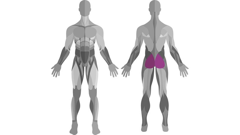


Gluteus Maximus
Shutterstock
What muscle is pictured here?
Teres Major
Gluteus Maximus
Holding the title of largest muscle in the human body, the gluteus maximus essentially makes up the butt cheek. It's responsible for helping you stand upright and keep posture. It also protects your bones when you sit.
Brachialis
Finger

Advertisement

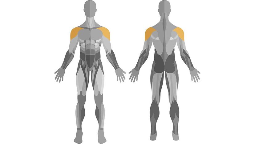
Deltoid
Shutterstock
What muscle is pictured here?
Deltoid
This muscle covers the shoulder joint - or glenohumeral joint - and has a rounded triangular shape, which explains why your shoulder is shaped the way it is. This muscle is named after the Greek alphabet letter delta, which is similar in shape to the muscle.
Pectoralis
Hammy
Tricep

Advertisement

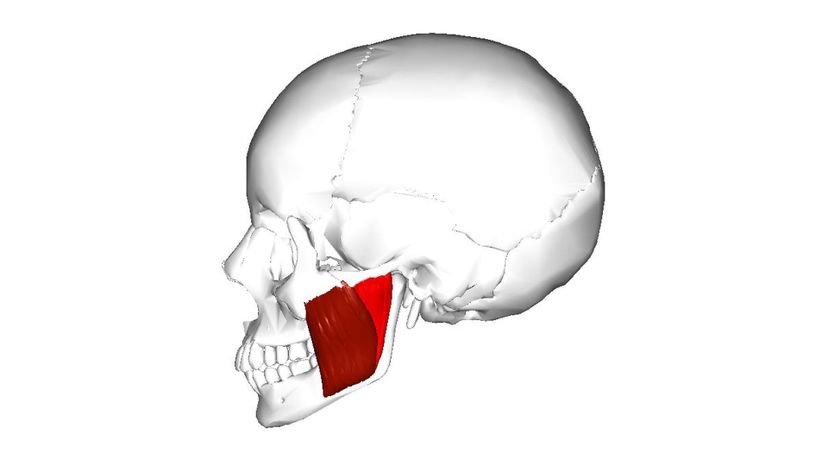
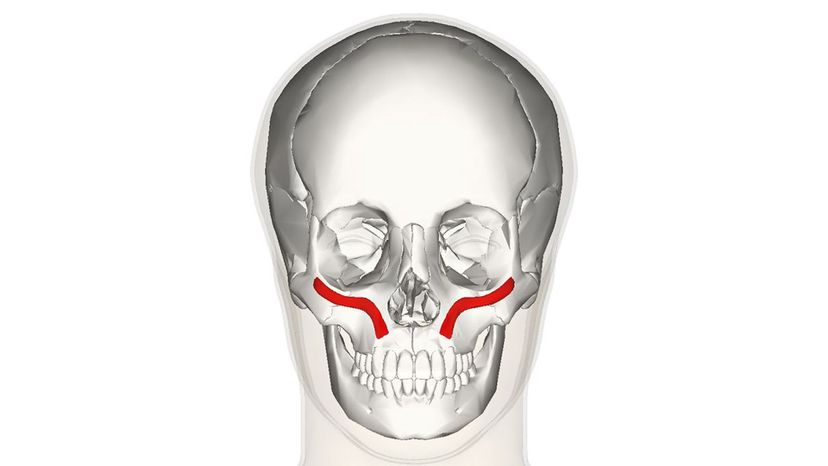
Masseter
Wiki Commons By Anatomography
What muscle is pictured here?
Intercostal muscles
Pronator Teres
Tendo Calcaneus
Masseter
Considering its small size, the masseter is a heavy lifter. Proportionally, it's the strongest muscle, capable of chomping down with a pressure up to 200 pounds when working together with the other jaw muscles.

Advertisement

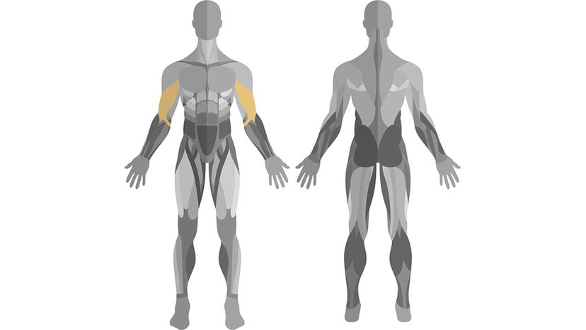
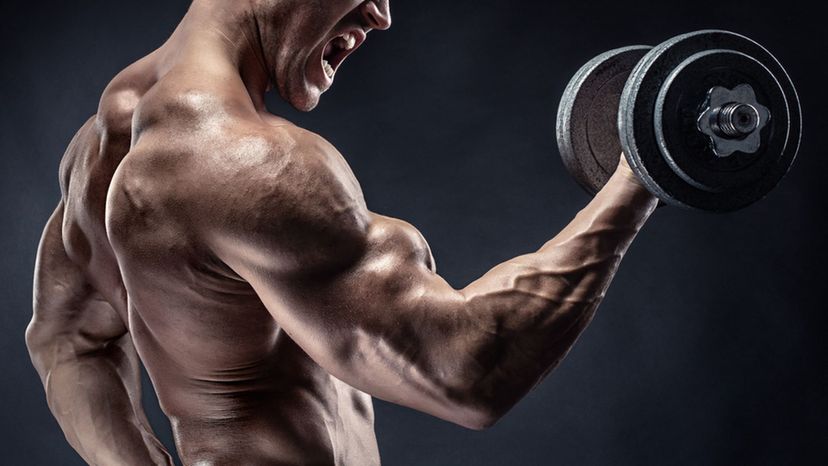
Biceps
Shutterstock
What muscle is pictured here?
Quadricep
Bicep
When you pull your arm up into a 90-degree angle and flex, that little bump that pops up is your bicep. It's possible to tear the bicep as well as develop tendinitis from overuse.
Sternum
Tibialus

Advertisement

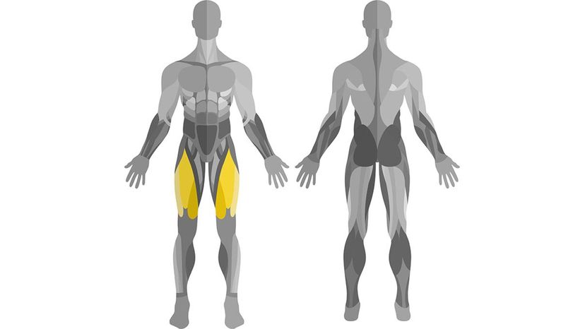
Quadriceps
Shutterstock
What muscle is pictured here?
Legeroni
Hammstring
Abdominus
Quadricep
The quadricep is a muscle group made of four components: vastus medialis, rectus femoris, vastus lateralis, and vastus intermedius. The group covers the front and sides of the upper leg.

Advertisement

Sternocleidomastoid
Shutterstock
What muscle is pictured here?
Hamstring
Frontalis
Serratus Anterior
Sternocleidomastoid
If you've ever irritated your sternocleidomastoid muscle, then turning your head from side-to-side must have been a literal "pain in the neck." This muscle runs from behind the ear at the base of the skull down to thorax (the center area of the collarbone and below) and allows you to turn your head from side to side, pull your chin to your chest, as well as tilt your head.

Advertisement

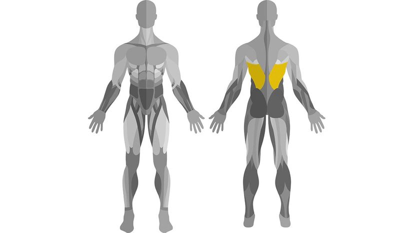
Latissimus Dorsi
Shutterstock
What muscle is pictured here?
Frontalis
Tricep
Latissimus Dorsi
Latissimus dorsi translates to "the broadest muscle of the back," a fitting name, considering it is one of the widest muscles in the body. Health enthusiasts and weight lifters usually refer to this muscle as the "lat."
Occipitalis

Advertisement

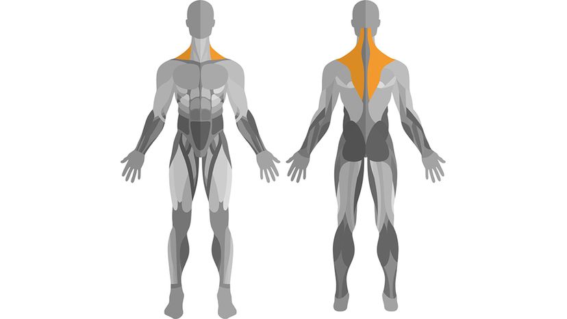
Trapizius
Shutterstock
What muscle is pictured here?
Ulnaris
Trapezius
The trapezius muscle runs from the base of the skull along the spine down to the lower inner tip of the shoulder blade on each side of the body. The muscle provides stabilization and allows the shoulder blade to move and rotate.
Deltoid
Hamstring

Advertisement

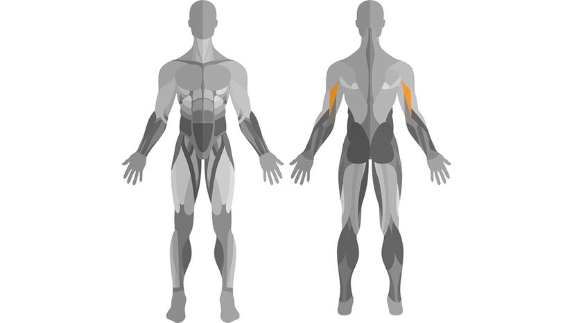
Tricep
Shutterstock
What muscle is pictured here?
Tricep
The tricep is one of the muscles in the back side of the upper arm. It allows the elbow to extend and retract.
Bicep
Deltoid
Magnep

Advertisement

Rectus Abdominis
Shutterstock
What muscle is pictured here?
Teres
Frontalis
Gracilis
Rectus Abdominus
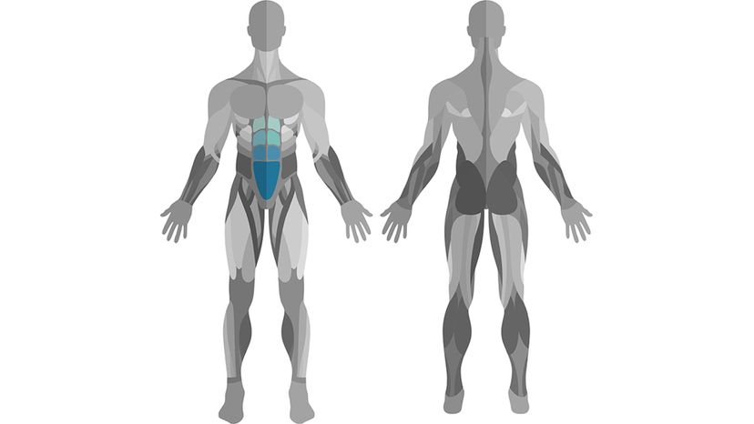
When people, especially men, talk about getting their midsections ready for bathing suit season, they're usually referring to their rectus abdominus, or abs. This muscle ranges from the pubic bone to the sternum. The abdominal muscles allow you to flex and move your midsection as well as greatly assist in balance and staying upright.​

Advertisement

Oblique (external internal)
Shutterstock
What muscle is pictured here?
Oblique
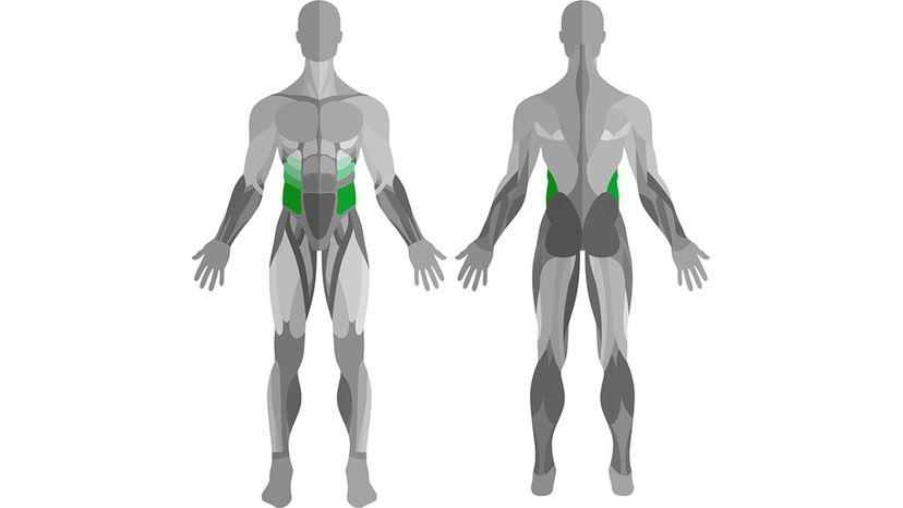
The external obliques run along your sides from the lower half of the ribs to the pelvis. These muscles primarily provide support in rotating the trunk of the body, but are also helpful when it comes to compressing the abdominal cavity and rotating the spine.
Trapezius
Lilac Moon
Thenar

Advertisement

Pectoralis Major
Shutterstock
What muscle is pictured here?
Tibialis
Bicep
Thenar
Pectoralis Major
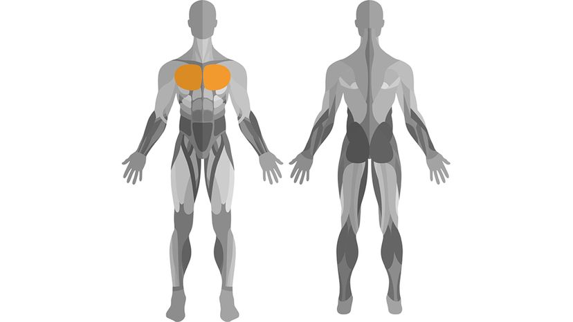
The pectoralis major muscle ranges from the breastbone to the shoulder and extends down around seven ribs. These breast muscles are commonly referred to as the "pecs."

Advertisement

Frontalis
Wikimedia Commons By Anatomography
What muscle is pictured here?
Ilioposoa
Hamstring
Frontalis
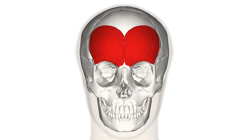
This muscle spans your forehead and makes many facial expressions possible. The frontalis muscle allows you to raise and furrow your brow. It's made of skeletal muscle, but it is not attached to any bones.
Serratus

Advertisement

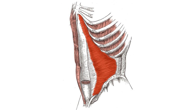
Transversus Abdominis
Wikimedia Commons By modified by Uwe Gille
What muscle is pictured here?
Temoralis
Transverse Abdominis
This muscle is also commonly known as the transversus abdominis or transversalis muscle. It makes up the innermost layer of abdominal muscles. It's key in protecting and holding your organs in place.
Oris
Mentalis

Advertisement

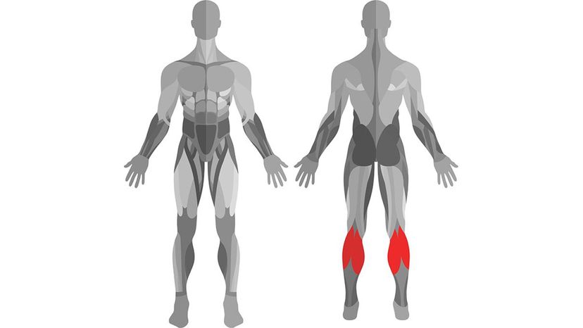
Gastrocnemius
Shutterstock
What muscle is pictured here?
Pronator Teres
Splenius Capitis
Gastrocnemius
This muscle works alongside the soleus muscle to make up the calf. Both muscles attach to the Achilles tendon as well as behind the knee. They also both run the entire length of the calf.
Tendo Calcaneus

Advertisement

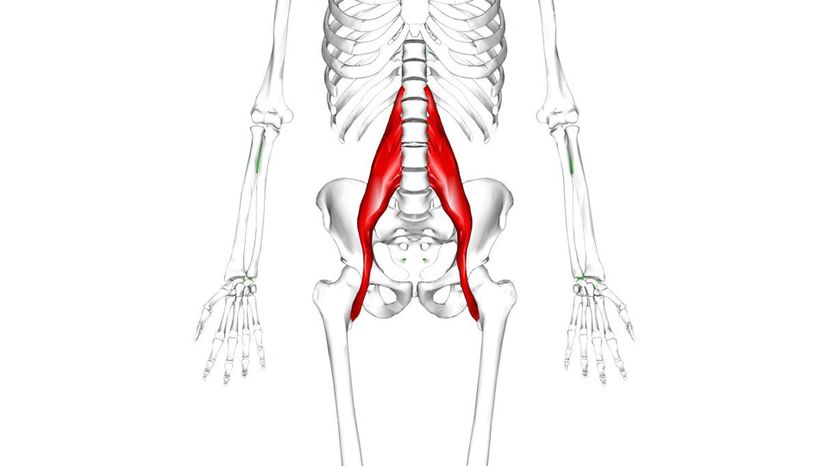
Iliopsoa
Wikimedia Commons By Anatomography
What muscle is pictured here?
Brachialis
Iliopsoas
The Iliopsoa muscle is made of the iliacus and psoas muscles in the pelvis. The two muscles are often clumped together because they are so similar in appearance and purpose.
Dermatitis
Biceps

Advertisement

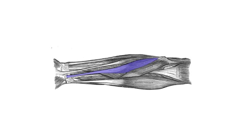
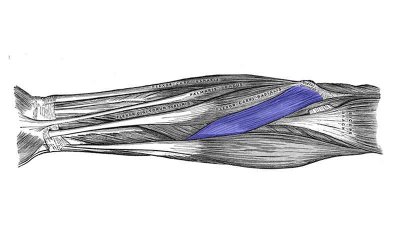
Flexor Carpi Radialis
Wikimedia Commons By Henry Gray
What muscle is pictured here?
Flexor Carpi Radialis
The flexor carpi radialis is the muscle that runs the length of the forearm. It attaches at the elbow and the base of the pointer finger and receives its blood supply from the ulnar artery. This muscle allows you to flex your wrist.
Frontalis
Anterior
Longus

Advertisement

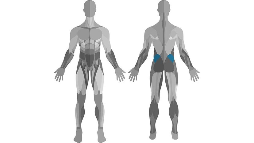
Gluteus Medius
Shutterstock
What muscle is pictured here?
Pronator
Tibialis
Gluteus Medius
The gluteus medius is essentially the side of the butt cheek, which helps in moving and rotating the thigh. This muscle also supports the pelvis for walking. It receives its blood supply from the superior gluteal artery. Damage or injury to the gluteus medius might result in an abnormal gait or limp.
Latae

Advertisement

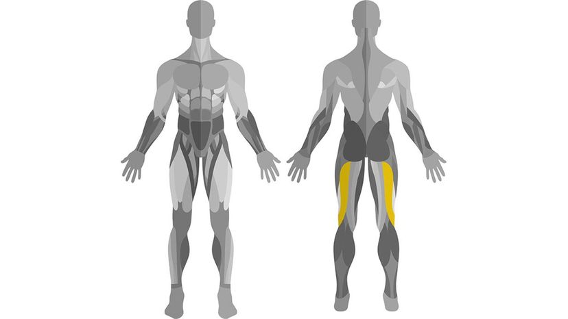
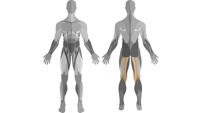
Hamstrings
Shutterstock
What muscle is pictured here?
Unaris
Biceps Femoris
The biceps femoris is one of three muscles of the hamstring group and is made up of two heads, a long one and a short one. The long one attaches at the lower back and hipbone and meets down at the femur above the knee, where it meets the short head and wraps around the knee and attaches to the fibula. The short head makes it possible for you to rotate your knee, while the long head helps with moving the leg and hip.
Masseter
Intercostal

Advertisement

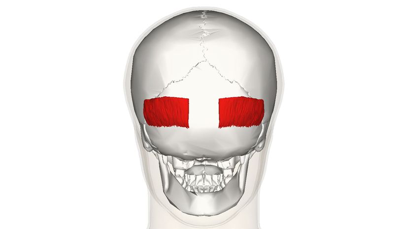
Occipitalis
Wikimedia Commons By Anatomography
What muscle is pictured here?
Splenius capitis
Occipitalis
This muscle is located near the occipital bone, which is at the base of the skull. The occipitalis muscle is part of a group of muscles that make up the epicranius muscle, which help make it possible for you to move your eyebrows and wrinkle your forehead.
Tendo calcaneus
Semitendinosus

Advertisement

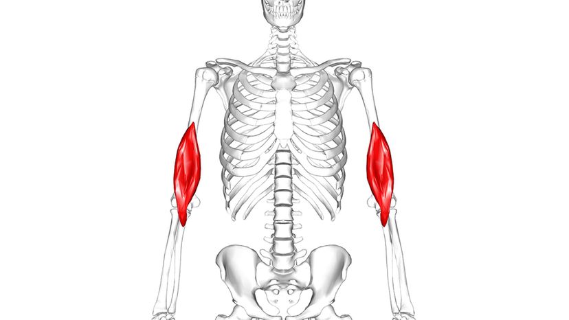
Brachialis
Wikimedia Commons By Anatomography
What muscle is pictured here?
Teres major
Brachialis
The brachialis lies deeper in the arm than the bicep and is a major muscle responsible for movement of the forearm. It's attached to the lower part of the humerus bone and receives its blood supply from two sources: the brachial and radial arteries.
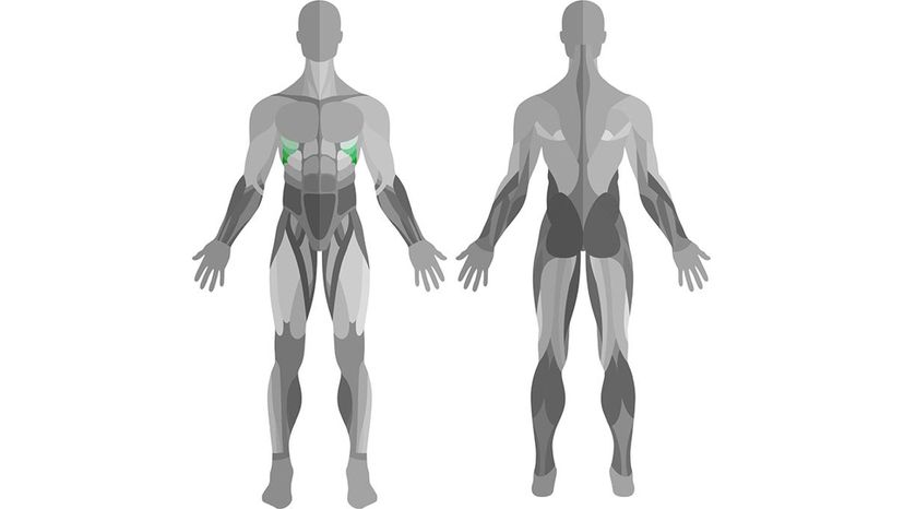
Infraspinatus
Trapezius

Advertisement

Intercostal muscles
Wikimedia Commons By Anatomography
What muscle is pictured here?
Levator Scapulae
Thenar
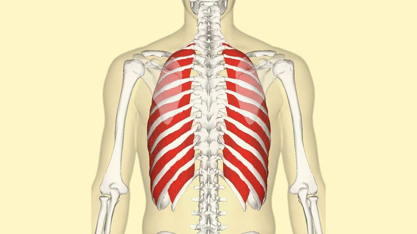
Intercostal muscles
There are 22 pairs of intercostal muscles, with 11 internal and 11 external ones. These muscles surround all the ribs, but the 12th is its bottom-most one. Intercostal muscles allow you to breathe.
Palmaris

Advertisement

Infraspinatus
Wikimedia Commons By Anatomography
What muscle is pictured here?
Femoral muscle
Semispinalis capitis
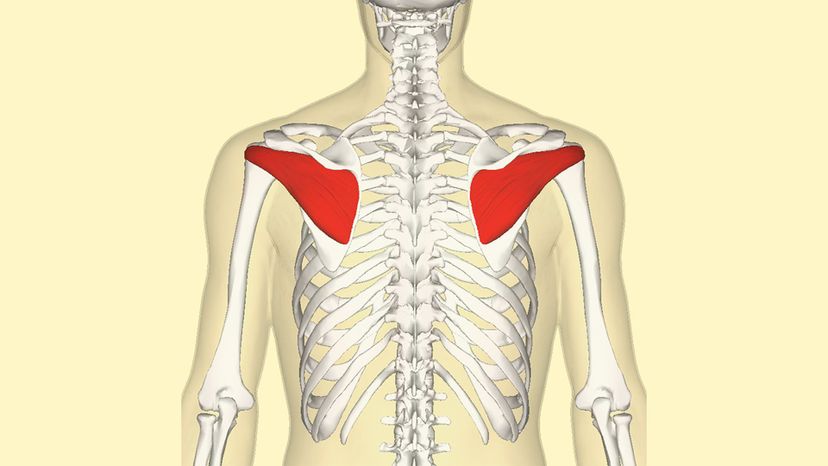
Infraspinatus
One of the four muscles that make up the rotator cuff, the infraspinatus is a triangular-shaped muscle that runs from the humerus across the shoulder blade. This muscle assists in stabilizing the shoulder joint and rotating the humerus.
Brachial teres

Advertisement

Teres Major
Shutterstock
What muscle is pictured here?
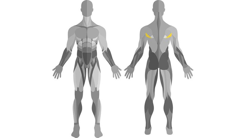
Teres major
The teres major muscle attaches to your lower shoulder blade while the teres minor muscle is found in​ the upper part of the shoulder blade. Both muscles then make their way over and insert into the humerus. They're relatively tiny muscles, smaller than the deltoid. The muscles' primary role is to assist in specific types of arm movement.
Gracilis
Fasciae Latae
Frontalis

Advertisement

Erector Spinae
Wikimedia Commons Henry Vandyke Carter
What muscle is pictured here?
Peroneus
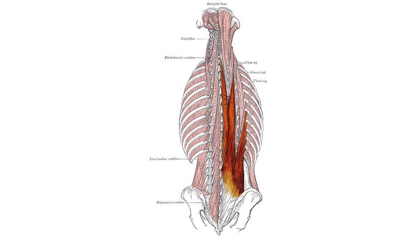
Erector Spinae
When it comes to rotating and straightening your back, you can thank the erector spinae muscle system found in the deep tissue of the back. It ranges the entire length of the back and has three parts.
Patellae
Tibialis

Advertisement

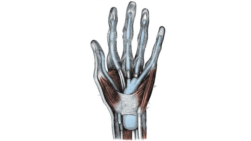
Thenar
Wikimedia Commons Henry Vandyke Carter
What muscle is pictured here?
Pronator Teres
Levator Scapulae
Thenar
The thenar grouping of muscles involves the thumb and includes the abductor pollicis muscle, the flexor pollicis brevis muscle, and the opponens pollicis deep muscle. On the other side of the hand is the hypothenar muscle group, which includes the abductor digiti minimi muscle, the flexor digiti minimi brevis muscle, and the opponens digiti minimi deep muscle.
Palmaris Longus

Advertisement

Semitendinosus
Shutterstock
What muscle is pictured here?
Semitendinosus
One of the three muscles that make up the hamstring, the semitendinosus muscle runs opposite to the biceps femoris (another hamstring muscle). It helps with rotating the tibia, especially when the knee is flexed, and also assists in flexing the knee and extending the upper leg or thigh.
Sternohyoid
Serratus Anterior
Tensor Fasciae Latae

Advertisement

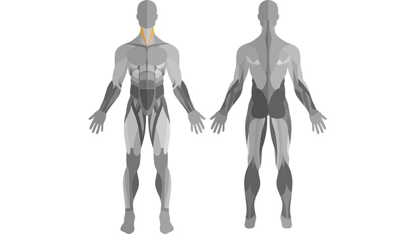
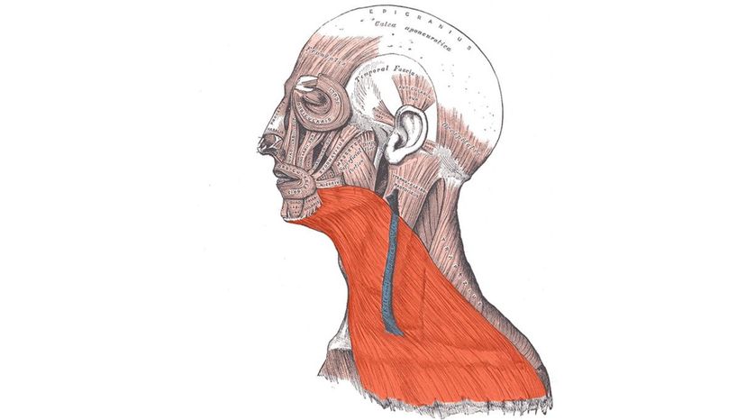
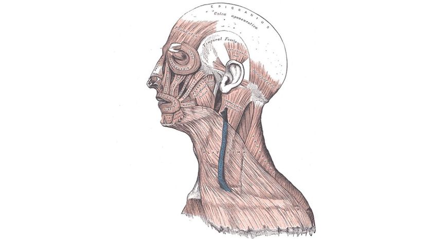
Platysma
Wiki Commons By modified by Uwe Gille
What muscle is pictured here?
Erector Spinae
Flexor Carpi Radialis
Transversus Abdominis
Platysma
The platysma is a layer of muscle fibers that run from the jawline down to the collar bone on the sides of the neck. If you want to stretch this muscle, sit or stand with relaxed shoulders and place your lower lip over your upper lip. Then, tilt your head back and you should feel a slight tug as the platysma muscle stretches.

Advertisement

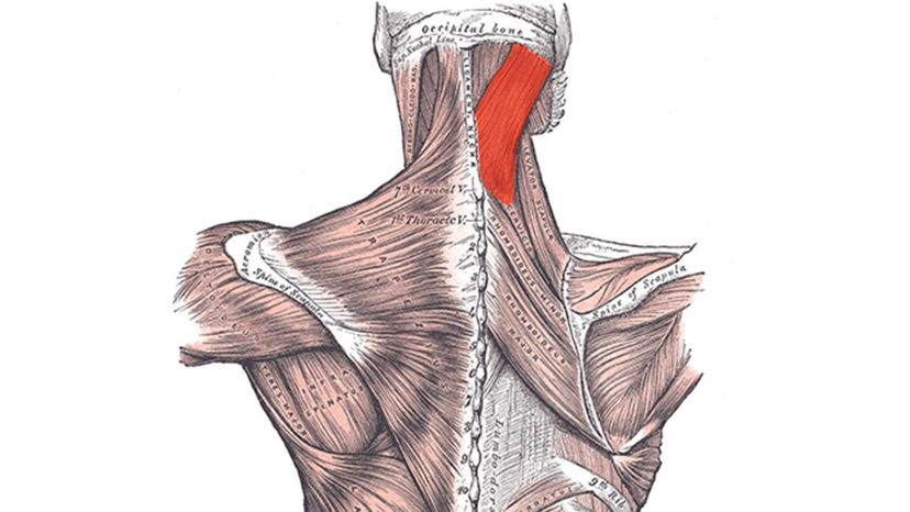
Splenius Capitis
Wikimedia Commons By modified by Uwe Gille
What muscle is pictured here?
Medius
Intercostal
Gracilis
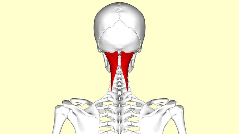
Splenius Capitis
The splenius capitis muscle forms a v-like shape at the back of the neck and connects the base of the skull to the upper spine. The muscles allow you to bend your head to the side as well as straighten it out.

Advertisement

Serratus Anterior
Shutterstock
What muscle is pictured here?
Cranial
Hamstring
Serratus Anterior
This muscle is located along the surface of the top eight ribs and allows for lifting the arms over the head. It also serves to protect the shoulder blade and receives its blood supply from the circumflex scapular artery.
Trapezius

Advertisement

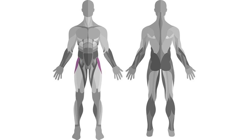
Gracilis
Shutterstock
What muscle is pictured here?
Semispinalis Capitis
Tibialis Anterior
Frontalis Superior
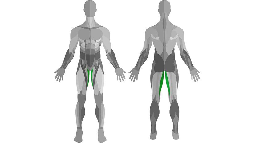
Gracilis
1 of five groin muscles, the gracilis muscle runs from the pubic bone all the way down to the upper part of the tibia (shinbone). It helps you stabilize and flex the knee as well as assists in hip adduction, which means the muscle is pulling a muscle towards the body rather than extending it away from the body.

Advertisement

Semispinalis Capitis
Wikimedia Commons By Anatomography
What muscle is pictured here?
Masseter
Semispinalis Capitis
Out of all the muscle layers in the transversospinalis muscle group, the semispinalis muscle is the most superficial. The transversospinalis muscle group is found in the deep tissue along the spine. This particular layer of muscle helps you keep your posture as well as assists in moving the spinal column and head.
hyoid
Tibialis Longus

Advertisement

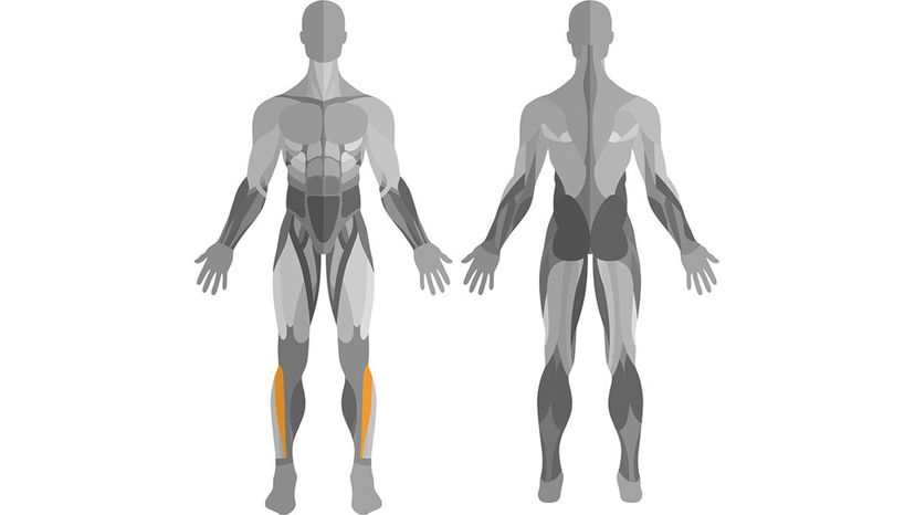
Extensor Digitorum Longus
Wikimedia Commons By modified by Uwe Gille
What muscle is pictured here?
Brachialis
Infraspinatus
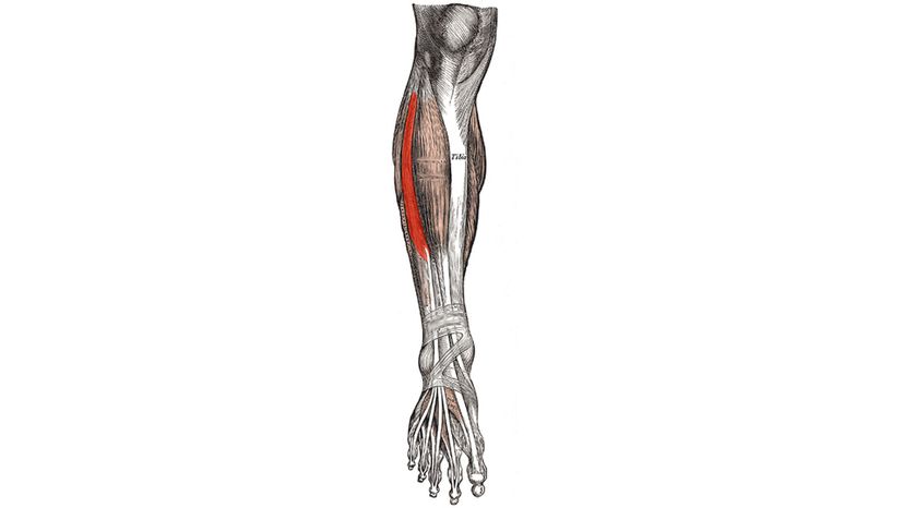
Extensor Digitorum Longus
This muscle begins at the upper part of the fibula and makes its way down the leg to the foot, where it divides to provide support to 4 of the 5 toes. However, it's not abnormal for some people to be born with Extensor Digitorum Longus muscle connections to the big toe as well.
Iliac Crest

Advertisement

Tibialis Anterior
Shutterstock
What muscle is pictured here?
Tibialis Anterior
This muscle attaches near the upper tibia and runs down to just above the ankle, where it turns into the tibialis tendon and continues on into the foot. The muscle allows you to pull your foot towards your shin, which is a vital part to balanced walking.
Pronator Teres
Levator
Crestoid

Advertisement

Tensor Fasciae Latae
Shutterstock
What muscle is pictured here?
Tensor Fasciae Latae
This muscle is found next the hip and butt cheek area on the side of the upper thigh. The muscle helps in stabilizing the hip and knee.
Frontalis
Femuricus
Mitochondriasis

Advertisement

Pronator Teres
Wikimedia Commons By Henry Gray
What muscle is pictured here?
Subcranial
Endocrine
Pronator Teres
This muscle is responsible for rotating the lower arm as well as flexing the forearm. It runs from the outer side of the elbow (Ulna) across to the radius.
Spenius Capitis

Advertisement

Occipitofrontalis
Wikimedia Commons Henry Vandyke Carter
What muscle is pictured here?
Palmaris Longus
Occipitofrontalis
The occipitofrontalis muscle includes the occipitalis muscle and the frontalis muscle. These muscles are part of the epicranial muscle layer that allows you to raise your eyebrows and wrinkle your forehead.
Levator Scapulae
Flexor Carpi Ulnaris

Advertisement

Sternohyoid
https://commons.wikimedia.org/wiki/File%3ASternohyoideus.png
What muscle is pictured here?
Carpi
Dorsi
Soleus
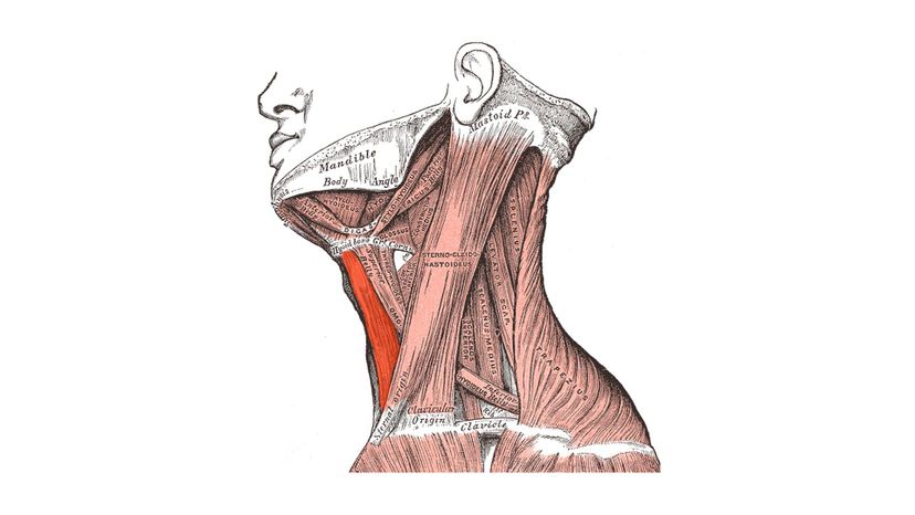
Sternohyoid
This petite, narrow muscle runs the entire length of the front of the neck and attaches the sternum to the hyoid bone. The sternohyoid contributes to speech and head and neck movement.

Advertisement

Levator Scapulae
Wikimedia Commons By Anatomography
What muscle is pictured here?
Iliac crest
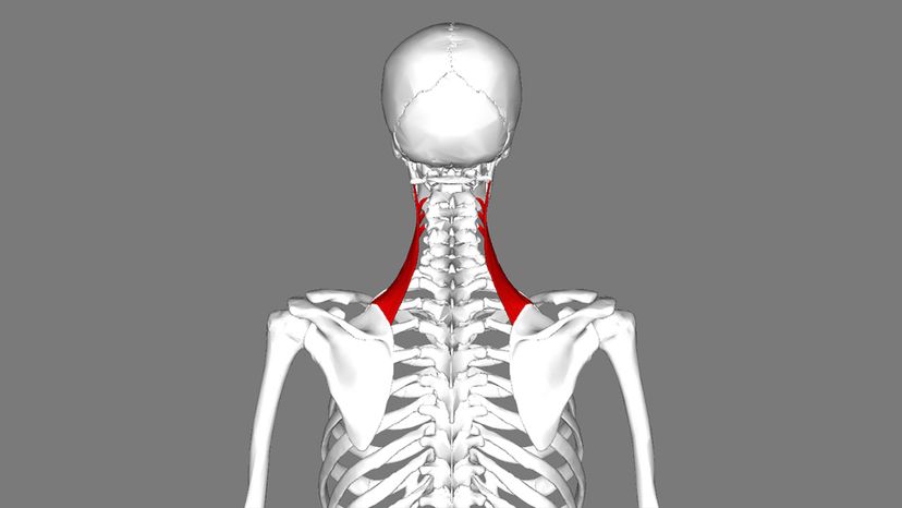
Levator Scapulae
This muscle attaches to the C1 through C4 vertebrae located in the upper neck, and then runs down to the top inner corner of the shoulder blade. This muscle allows you to shrug your shoulders and is partially covered by the sternocleidomastoid and trapezius muscles.
Palmaris Longus
Splenius Capitis

Advertisement

Quadratus Femoris
Wikimedia Commons By Mikael Häggström
What muscle is pictured here?
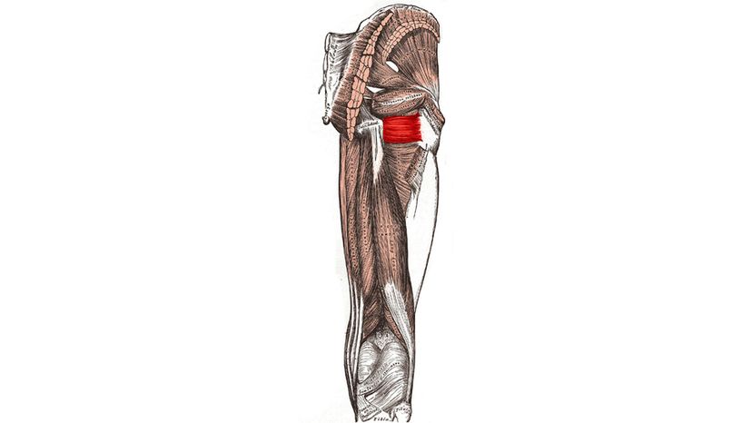
Quadratus Femoris
To stretch the quadratus femoris muscle, you'll have to contort yourself into the standing pigeon position. To do this, place your feet under your hips and then cross your left foot over your right ankle. Bend your right knee into a squatting position, but ensure your knee does not jut out past your toes at any point. Try to lean into the stretch so that your forearms are on your left shin.
Tricep
Maximus
Brachialis

Advertisement

Diaphram
Youtube
What muscle is pictured here?
Intestine
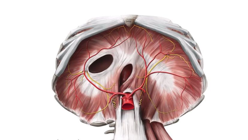
Diaphram
The diaphragm is made of skeletal muscle. It allows you to breathe by flexing and relaxing, which draws in and exhales air. The muscle structure also serves as a barrier between the thoracic and abdominal cavities.
Trapezius
Tricep

Advertisement

Zygomaticus Minor
Wikimedia Commons By Anatomography
What muscle is pictured here?
Zygomaticus Minor
Similar to the zygomaticus major, the zygomaticus minor is involved in moving the mouth. However, while its counterpart assists in smiling, the minor muscle makes frowning possible.
Bicep
Ilioposoa
Gastrocnemius

Advertisement

You Got:
/42
Shutterstock